
Coarctation of the aorta
Conditions
Overview
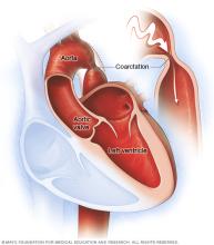
Aortic coarctation (ko-ahrk-TAY-shun) is a narrowing of part of the aorta. The aorta is the main artery of the body. Aortic coarctation also is called coarctation of the aorta.
The condition forces the heart to work harder to pump blood. It usually is present at birth. That means it is a congenital heart defect. But sometimes the condition can occur later in life.
Coarctation of the aorta often occurs along with other congenital heart defects. Treatment to fix the condition is usually successful. But regular health checkups are needed for life to watch for changes in the heart's health.
Symptoms
Symptoms of coarctation of the aorta depend on how much of the aorta is narrowed. Most people don't have symptoms.
Adults and older children with mild coarctation of the aorta may not have symptoms. Their hearts may seem healthy.
If a baby is born with an extreme narrowing of the aorta, symptoms may be seen shortly after birth. Symptoms of coarctation of the aorta in infants include:
- Trouble breathing.
- Trouble feeding.
- Heavy sweating.
- Irritability.
- Blue or gray skin, lips and fingernails. Depending on the skin color, these changes may be harder or easier to see.
Symptoms of coarctation of the aorta later in life may include:
- Chest pain.
- High blood pressure.
- Headaches.
- Muscle weakness.
- Leg cramps.
- Cold feet.
- Nosebleeds.
Coarctation of the aorta often occurs with other heart conditions present at birth. Other symptoms depend on the types of congenital heart defects.
When to see a doctor
Always get medical help right away for chest pain that is sudden or not explained.
Also get medical help for these symptoms:
- Fainting.
- Sudden shortness of breath.
- High blood pressure that can't be explained.
Many different health conditions can cause these symptoms. A complete health checkup is needed to learn the cause.
Causes
The cause of coarctation of the aorta is not clear. It's usually a heart condition present at birth, called a congenital heart defect. A congenital heart defect happens as the baby, called a fetus, is growing in the womb during pregnancy. The cause is often not known.
Rarely, coarctation of the aorta can happen later in life. Conditions or events that can narrow the aorta and cause this condition include:
- Traumatic injury.
- A buildup of fats, cholesterol and other substances in and on the artery walls, called atherosclerosis.
- A rare type of swelling and irritation of blood vessels in the heart, called Takayasu arteritis.
Risk factors
Risk factors for coarctation of the aorta include:
- Male sex.
- Some genetic conditions, such as Turner syndrome.
- Some heart conditions present at birth, called congenital heart defects.
Congenital heart defects that can occur with coarctation of the aorta include:
- Bicuspid aortic valve. The aortic valve is between the lower left chamber of the heart and the aorta, which is the body's main artery. Typically, the aortic valve has three cusps. A bicuspid aortic valve has only two cusps.
- Subaortic stenosis. This is a narrowing of the area below the aortic valve. It blocks blood flow from the lower left heart chamber to the aorta.
- Patent ductus arteriosus. The ductus arteriosus is a blood vessel that connects the lung arteries to the aorta. When a baby is growing in the womb, this vessel lets blood go around the lungs. Shortly after birth, the ductus arteriosus usually closes. If it stays open, it's called a patent ductus arteriosus.
- Holes in the heart. Some people with coarctation of the aorta also are born with a hole in the heart. If the hole is between the upper heart chambers, it is called an atrial septal defect. A hole between the lower heart chambers is called a ventricular septal defect.
- Congenital mitral valve stenosis. This is a type of heart valve disease that some people are born with. The valve between the upper and lower left heart chambers is narrowed or small. It's harder for blood to move through the valve.
Complications
Complications of coarctation of the aorta happen because the lower left heart chamber has to work harder to pump blood through the narrowed artery. This makes blood pressure go up in the lower left heart chamber. Also, the wall of the chamber can get thick. This condition is called ventricular hypertrophy.
Complications of coarctation of the aorta include:
- A bulge in the wall of the aorta, called an aortic aneurysm.
- A bulge or ballooning in a blood vessel in the brain, also known as a brain aneurysm.
- A rupture or tear in the aorta, called an aortic dissection.
- Bleeding in the brain.
- Coronary artery disease.
- High blood pressure that doesn't go away. Blood pressure usually drops after surgery to fix the aorta, but it may still be higher than usual.
- Stroke.
Treatment may be needed right away to prevent complications. Without treatment, coarctation of the aorta may lead to:
- Kidney failure.
- Heart failure.
- Death.
Some people have complications after treatment for coarctation of the aorta. These complications include:
- Repeat narrowing of the aorta, called recoarctation.
- Aortic aneurysm or rupture.
To prevent complications, people with coarctation of the aorta need regular health checkups for life.
Prevention
There's no known way to prevent coarctation of the aorta. Tell your healthcare team if anyone in your family was born with a heart condition.
Diagnosis
Diagnosis of coarctation of the aorta may depend on how severe the heart condition is. Severe coarctation of the aorta is usually found soon after birth. The condition may be seen on ultrasound images taken during pregnancy.
If the condition is mild, it may not be found until later in life.
To diagnose coarctation of the aorta, a healthcare professional checks blood pressure in the arms and legs. Depending on which part of the aorta is affected, blood pressure may be high in the arms and low in the legs and ankles. The pulse may be weaker or slower to feel in the legs compared to the arms.
A whooshing sound called a heart murmur may be heard when listening to the heart.
Tests
Tests to diagnose coarctation of the aorta may include:
- Echocardiogram. Sound waves make pictures of the beating heart. An echocardiogram shows the structure of the heart chambers and valves. It also shows how well blood moves through the heart and heart valves. The test can often tell which part and how much of the aorta is narrowed. An echocardiogram also helps healthcare professionals plan treatment for coarctation of the aorta.
- Chest X-ray. A chest X-ray shows the condition of the heart and lungs. A chest X-ray might show a narrowing in the aorta where the coarctation is.
- Cardiac magnetic resonance imaging (MRI). This test uses magnetic fields and radio waves to create detailed images of the heart and blood vessels. It can show which part and how much of the aorta is narrowed. A healthcare professional also may use MRI results to guide treatment.
- Computerized tomography (CT) scan of the heart. This test uses several X-rays to make detailed pictures of the heart. It may be used if other tests don't give enough information to make a diagnosis.
-
Cardiac catheterization with angiogram. An angiogram uses X-rays to look at the heart's blood vessels, including the aorta. It's usually done to see if a blood vessel is narrowed or blocked. An angiogram is sometimes done as part of a general group of heart tests and treatments called cardiac catheterization.
During cardiac catheterization, a doctor places a thin flexible tube called a catheter into a blood vessel, usually in the groin or wrist. The doctor guides the tube to the heart. A substance called contrast flows through the tube to arteries in the heart. The contrast makes the arteries easier to see on pictures taken during the test. Cardiac catheterization can help show how much of the aorta is narrowed. The doctor also can measure the pressure in your heart and blood vessels.
- CT angiogram. This test looks at the arteries that supply blood to the heart. It uses a powerful X-ray machine to make images of the heart and its blood vessels. A CT angiogram uses contrast to show how blood flows through the veins and arteries. The test can show where and how severe coarctation of the aorta is. It also can tell whether other blood vessels are affected. A CT angiogram also can be used to guide treatment.
Treatment
Treatment for coarctation of the aorta depends on how old the person is when the condition is diagnosed. Treatment also depends on how much of the aorta is narrowed.
Coarctation of the aorta treatment may include:
- Medicines.
- A heart procedure.
- Surgery.
If there are other congenital heart defects, they may be fixed at the same time.
Medication
Medicines for coarctation of the aorta may include:
- Blood pressure medicines. Medicines are used to control blood pressure before repair surgery. Although fixing the aorta can improve blood pressure, many people still need blood pressure medicine after the repair surgery.
- Medicine to keep the ductus arteriosus open. Babies are born with a temporary opening called the ductus arteriosus between the aorta and the pulmonary artery. This opening lets some blood go to the lungs. The ductus arteriosus most often closes soon after birth. But medicine can keep it open. Babies with severe coarctation of the aorta often get such medicine until surgery is done to fix the aorta.
Surgery or other procedures
Surgery or a heart procedure can be done to repair coarctation of the aorta. Options include:
-
Balloon angioplasty and stenting. This may be the first treatment for coarctation of the aorta. Sometimes it's done if the aorta gets narrow again after coarctation surgery. The treatment helps widen a narrowed artery and improve blood flow.
During angioplasty, a doctor uses a thin tube called a catheter and a tiny balloon to open a narrowed artery. Usually, a small metal mesh tube called a stent is placed in the artery. The stent keeps the artery open. It also lowers the risk of the artery getting narrow again.
- Resection with end-to-end anastomosis. A surgeon takes out the narrowed part of the aorta. This is called a resection. The surgeon then connects the two healthy parts of the aorta. This is called anastomosis.
- Bypass graft repair. This surgery uses a tube called a graft to create a new path for blood to flow around the narrowed area of the aorta.
- Patch aortoplasty. The surgeon cuts across the narrowed area of the aorta. Then the surgeon patches in a piece of material to widen the narrowed part of the blood vessel. This treatment is useful if the coarctation involves a long part of the aorta.
After aortic repair surgery, health checkups are needed for life to keep track of blood pressure and watch for complications.
Lifestyle and home remedies
If you had coarctation of the aorta, it's important to take steps to manage blood pressure and keep the heart healthy.
Try these heart-healthy steps:
- Do not smoke or use tobacco. If you need help quitting, talk with your healthcare team.
- Eat healthy foods. Choose plenty of fruits, vegetables and whole grains. Limit sugar, salt, and trans and saturated fats.
- Exercise and stay active. Talk with your healthcare team about the amount and type of exercise that's right for you. Some physical activities, such as weightlifting, can temporarily raise blood pressure.
- Manage weight. Ask your healthcare team what a healthy weight is for you.
- Manage blood pressure, cholesterol and diabetes. Ask your healthcare team how often you need checkups.
- Lower and manage stress. Some tips are to get more exercise, practice mindfulness and connect with others in support groups.
- Get good sleep. Adults should try to get 7 to 9 hours of sleep daily.
Also talk with your healthcare team about these things:
- Pregnancy. Coarctation of the aorta, even after it's fixed, may raise the risk of an aortic tear or rupture during pregnancy and childbirth. Before becoming pregnant, talk with a doctor trained in congenital heart diseases, called an adult congenital cardiologist. Together you can go over the possible risks and complications.
- Antibiotics before dental work. Bacteria can affect the inner lining of the heart or valves, causing an infection called endocarditis. People with some heart conditions may need to take antibiotics before dental work to prevent this infection. Ask your healthcare professional whether you need preventive antibiotics.
Preparing for an appointment
If coarctation of the aorta is diagnosed at birth, there may not be time to prepare. If symptoms aren't noticed until later in life, see a doctor trained in heart conditions present at birth. This type of healthcare professional is called a congenital cardiologist.
Here's some information to help you prepare for the appointment.
What you can do
Write down the following and bring the notes with you to your appointment:
- Any symptoms, even if they don't seem related to your heart. Include when each symptom began.
- Important medical information, including any family history of heart conditions present at birth.
- All medicines, vitamins and supplements you take and the doses. Include those bought without a prescription. Also tell your healthcare professional about any taken during pregnancy.
- Any questions you want to ask your healthcare professional.
Questions to ask the healthcare professional include:
- How is coarctation of the aorta treated?
- What treatment do you recommend and why?
- What is recovery and rehabilitation like after my treatment or surgery?
- How often do I need to follow-up tests and checkups?
- What is the risk of complications from this condition?
- What symptoms or warning signs should I watch for?
- What is the long-term outlook?
- How should I change my diet or exercise and sports activities?
- Are antibiotics needed before dental work or other medical procedures?
- Is pregnancy safe for those with coarctation of the aorta?
- What is the risk that my or my child's future children will have coarctation of the aorta?
Don't hesitate to ask any other questions.
Also ask someone to go with you to the appointment, if possible. Someone who goes with you can help remember what the health professional says.
What to expect from your doctor
Members of your healthcare team are likely to ask you many questions if they think your child has coarctation of the aorta. Being ready to answer them may save time to go over anything you want to spend more time on. Your healthcare team may ask:
- When did you first notice your child's symptoms?
- Can you describe your child's symptoms?
- Does your child have any breathing trouble, such as fast breathing or running out of breath easily?
- Does your child tire easily?
- Does your child sweat heavily?
- Does your child seem irritable?
- Does your child have chest pain?
- Does your child often have cold feet?
- When do these symptoms happen?
- Do the symptoms come and go, or does your child always have them?
- Do the symptoms seem to be getting worse?
- Do you have a family history of congenital heart defects?
- What, if anything, makes your child's symptoms better?
- What, if anything, makes your child's symptoms worse?
If you had coarctation of the aorta, your healthcare team may ask:
- When did the symptoms start?
- Have the symptoms gotten worse over time?
- Does exercise or activity make your symptoms worse?
- Have you been diagnosed with any other medical conditions?
- Do you have a family history of heart conditions?
- Are you planning to become pregnant in the future?
© 1998-2026 Mayo Foundation for Medical Education and Research(MFMER). All rights reserved. Terms of Use