CT scan
Procedures
Overview

A computerized tomography scan, also called a CT scan, is a type of imaging that uses X-ray techniques to create detailed images of the body. It then uses a computer to create cross-sectional images, also called slices, of the bones, blood vessels and soft tissues inside the body. CT scan images show more detail than plain X-rays do.
A CT scan has many uses. It's used to diagnose disease or injury as well as to plan medical, surgical or radiation treatment.
Why it's done
Your healthcare professional may suggest a CT scan for many reasons. For instance, a CT scan can help:
- Diagnose muscle and bone conditions, such as bone tumors and breaks, also called fractures.
- Show where a tumor, infection or blood clot is.
- Guide procedures such as surgery, biopsy and radiation therapy.
- Find and watch the progress of diseases and conditions such as cancer, heart disease, lung nodules and liver masses.
- Watch how well certain treatments, such as cancer treatment, work.
- Find injuries and bleeding inside the body that can happen after trauma.
Risks
Radiation exposure
During a CT scan, you're briefly exposed to a type of energy called ionizing radiation. The amount of radiation is greater than the amount from a plain X-ray because the CT scan gathers more-detailed information.
The low doses of radiation used in CT scans have not been shown to cause long-term harm. But for repeated scans, there may be a small increase in the lifetime risk of cancer. This can affect children more than adults.
CT scans have many benefits that outweigh any small risk. Healthcare professionals use the lowest dose of radiation to get the needed medical information. And newer, faster machines and techniques use less radiation than older CT scans did. Talk with your healthcare professional about the benefits and risks of a CT scan.
Harm to unborn babies
Tell your healthcare professional if you're pregnant. The radiation from a CT scan is unlikely to harm your baby unless the scan is of your belly or pelvis. But your health professional might suggest another type of exam so that the baby isn't exposed to radiation. Exams that don't use radiation include ultrasound and MRI.
Contrast material
A special dye called contrast material is needed for some CT scans. The dye appears bright on images. So it makes certain areas of the body that are being scanned show up better. This can help make blood vessels, intestines or other structures easier to see.
Contrast material might be given:
- By mouth. If your esophagus or stomach is being scanned, you may need to swallow a liquid that has contrast material. This drink may not taste good.
- By shot, also called injection. Contrast agents can be given through an artery or a vein in your arm. You may get a feeling of warmth or a metallic taste in your mouth when the dye goes into your body.
- By enema. A contrast material may be put in your rectum to show your intestines. This procedure can make you feel bloated.
Reactions to contrast material
Although rare, medical problems or allergic reactions can happen with contrast material. Most reactions are mild and result in a rash or itchiness. More rarely, an allergic reaction can be serious, even life-threatening. Tell your healthcare professional if you've ever had a reaction to contrast material.
How you prepare
Depending on which part of your body is being scanned, you may be asked to:
- Take off some or all your clothing and wear a hospital gown.
- Remove metal objects, such as belts, jewelry, dentures and eyeglasses, that might affect image results.
- Not eat or drink for a few hours before your scan.
Preparing your child for a scan
If your infant or toddler is having a CT scan, the healthcare professional may suggest a medicine called a sedative to help keep your child calm and still. Movement blurs the images and may affect the results. Ask your health professional how to help get your child ready for the scan.
What you can expect
You can have a CT scan in a hospital or an outpatient facility. CT scans are painless. With newer machines, scans take only a few minutes. The whole process most often takes about 30 minutes.
During the procedure
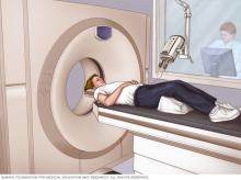
A CT scanner is shaped like a large doughnut standing on its side. You lie on a narrow table with a motor that slides through the center of the scanner into a tunnel. Straps and pillows may be used to help you stay in place. During a head scan, the table may be fitted with a special cradle that holds your head still.
While the table moves you into the scanner, the X-ray tube rotates around you. Each time it rotates, it gives images of thin slices of your body. You may hear buzzing and whirring noises.
A healthcare professional called a CT technologist sits in another room and can see and hear you. You can talk with the technologist through an intercom. To help you keep still during the scan, the technologist might ask you to hold your breath at certain points. Movement can blur the images.
After the procedure
After the exam you can return to your regular routine. If you were given contrast dye, you may be asked to wait for a short time before leaving to make sure that you feel OK after the exam. You also might be told to drink lots of fluids to help your kidneys remove the dye from your body.
Results
CT images are stored as electronic data files. They're most often reviewed on a computer screen. A doctor who specializes in imaging, called a radiologist, looks at the images and creates a report that's kept in your medical records. Your healthcare professional talks with you about the results.
© 1998-2026 Mayo Foundation for Medical Education and Research(MFMER). All rights reserved. Terms of Use
